Diagram Of Shoulder Joint : The Shoulder Joint / Describe the structure of the shoulder should begin with bone parts that include:. The glenohumeral joint is the main joint of the shoulder and the generic term shoulder joint usually refers to it. Muscle diagram of shoulder shoulder muscles diagram shoulder muscle diagrams diagram site. Here, we shall consider the factors the permit movement, and. Find & download the most popular shoulder joint vectors on freepik free for commercial use high quality images made for creative projects. Human shoulder diagram human shoulder anatomy stock photo anatomyinsider 129018944.
Equally extensive are the muscles affecting the shoulder movement, including: It can also be called abduction as the movement pulls the scapula away from the vertebrae. The shoulder joint may be subjected to different injuries. The glenohumeral joint is the main joint of the shoulder and the generic term shoulder joint usually refers to it. There are two kinds of cartilage in the joint.
Shoulder Anatomy Alila Medical Images from m.psecn.photoshelter.com It is formed by the head of the humerus and the glenoid cavity of the scapula. Atlas of the anatomy of the joint of the shoulder on a ct arthrogram in axial, coronal, and sagittal sections, on a 3d images and on conventional athrogram. Posted on november 17, 2018november 17, 2018. It is the major joint connecting the upper limb the shoulder joint is one of the most mobile in the body, at the expense of stability. Clavicle fracture with broken collarbone vector illustration. Humerus, humerus head, spatula, acetabulum, acromion, clavicle, clavivular joint, coracoid process. The glenohumeral joint is the main joint of the shoulder and the generic term shoulder joint usually refers to it. Diagram of the human shoulder joint.
Editor · aug 8, 2017 ·.
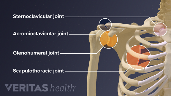
Cumpără planșa joints of the lower extremities anatomical chart la prețul de 50.06 lei, discount 5% cu livrare prin curier oriunde în românia. Nerve innervation of the shoulder joint. Diagram of the human shoulder joint. Human shoulder diagram human shoulder anatomy stock photo anatomyinsider 129018944. A selection of frequent lesions encountered by medical students as well as skilled orthopedists and surgeons, and neurologists, is presented here. This diagram here just shows the joint capsule itself. Posted on november 17, 2018november 17, 2018. Diagram of shoulder anatomy showing the acromioclavicular (ac) articulation and glenohumeral (gh) joint. Clinical examples of shoulder joint injuries or lesions. Atlas of the anatomy of the joint of the shoulder on a ct arthrogram in axial, coronal, and sagittal sections, on a 3d images and on conventional athrogram. Where the rounded top of the arm bone (humerus) contacts the shoulder blade is called the glenohumeral joint. Click now and learn everything about its anatomy and function at kenhub! The glenohumeral, or shoulder, joint is a synovial joint that attaches the upper limb to the axial skeleton.
Simple easy notes for quick revision for exams. The shoulder joint (glenohumeral joint) is a ball and socket joint between the scapula and the humerus. Find & download the most popular shoulder joint vectors on freepik free for commercial use high quality images made for creative projects. It is formed by the head of the humerus and the glenoid cavity of the scapula. Most relevant best selling latest uploads.
Guide To Shoulder Anatomy from embed.widencdn.net The glenohumeral or shoulder joint, is a ball and socket joint. Posted on november 17, 2018november 17, 2018. The shoulder joint may be subjected to different injuries. We can also call this adduction of the scapulae. protraction is the pulling forward of the shoulder joint. The left shoulder and acromioclavicular joints, and the proper ligaments of the scapula. Movement in this part of the body is more complex than in other large joints, such as the hip or knee. The first type is the white cartilage on the ends of the bones (called articular cartilage) which allows the bones to glide and move on each other. Suprascapular , axillary, subscapular , lateral pectoral and musculocutaneous nerve branches.
Nerve innervation of the shoulder joint.
Editor · aug 8, 2017 ·. The shoulder joint is vulnerable to dislocations from sudden jerks of the arm, especially in children before strong muscles have developed. Clinical examples of shoulder joint injuries or lesions. There are two kinds of cartilage in the joint. Following inferior dislocation of shoulder joint, the rounded contour of shoulder is lost and there is weakness of abduction of armbecause the. Where the rounded top of the arm bone (humerus) contacts the shoulder blade is called the glenohumeral joint. Learn vocabulary, terms and more with flashcards, games and other study tools. Just remember the articulating surfaces. Atlas of the anatomy of the joint of the shoulder on a ct arthrogram in axial, coronal, and sagittal sections, on a 3d images and on conventional athrogram. Most relevant best selling latest uploads. Shoulder joint anatomy and information the most flexible joint in the entire human body our shoulder joint is formed by the union of the humerus the scapula or shoulder blade and the shoulder muscles anatomy diagram & function. Equally extensive are the muscles affecting the shoulder movement, including: A selection of frequent lesions encountered by medical students as well as skilled orthopedists and surgeons, and neurologists, is presented here.
Simply put, the shoulder, or shoulder joint, is the connection of the upper arm and the thorax. We can also call this adduction of the scapulae. protraction is the pulling forward of the shoulder joint. Diagram of the human shoulder joint. Know every tiny but important part of your arms from the humeral head to the scapula. The shoulder joint (glenohumeral joint) is a ball and socket joint between the scapula and the humerus.
Shoulder Anatomy Biomechanics Proprofs Quiz from media.proprofs.com It can also be called abduction as the movement pulls the scapula away from the vertebrae. Human shoulder diagram human shoulder anatomy stock photo anatomyinsider 129018944. This is called the glenoid. Simply put, the shoulder, or shoulder joint, is the connection of the upper arm and the thorax. Nerve innervation of the shoulder joint. It is the major joint connecting the upper limb the shoulder joint is one of the most mobile in the body, at the expense of stability. It is formed by the head of the humerus and the glenoid cavity of the scapula. The shoulder joint is vulnerable to dislocations from sudden jerks of the arm, especially in children before strong muscles have developed.
Shoulder diagram illustrations & vectors.
The shoulder joint is supplied with blood by branches of the anterior and posterior circumflex humeral arteries, the suprascapular artery and the scapular circumflex diagram of the human shoulder joint, back view. Click now and learn everything about its anatomy and function at kenhub! Posted on november 17, 2018november 17, 2018. It is a ball and socket joint that allows the arm to rotate in a circular fashion or to hinge out. We can also call this adduction of the scapulae. protraction is the pulling forward of the shoulder joint. Shoulder diagram illustrations & vectors. A selection of frequent lesions encountered by medical students as well as skilled orthopedists and surgeons, and neurologists, is presented here. You can see it enclosing the glenohumeral joint and you can see its attachment on the anatomical neck that's the shoulder joint. Check out this shoulder joint science diagram template in the edraw free download resources library. Atlas of the anatomy of the joint of the shoulder on a ct arthrogram in axial, coronal, and sagittal sections, on a 3d images and on conventional athrogram. Human shoulder joint pain anatomy. Retraction pulls the shoulder joint to the rear and toward the vertebral column. Movement in this part of the body is more complex than in other large joints, such as the hip or knee.
Where the rounded top of the arm bone (humerus) contacts the shoulder blade is called the glenohumeral joint diagram of shoulder. The glenohumeral joint is the main joint of the shoulder and the generic term shoulder joint usually refers to it.


(125).jpg)


0 Comments