Foot Muscles Mri / MRI of the foot radiopedia - Anatomical structures of the ankle and foot and specific regions (major joints) are visible as dynamic labeled images.
Foot Muscles Mri / MRI of the foot radiopedia - Anatomical structures of the ankle and foot and specific regions (major joints) are visible as dynamic labeled images.. Muscle anatomy books free download 12 photos of the muscle anatomy books free download muscle anatomy books free download, human muscles, muscle anatomy books free download Both muscles are innervated by the deep fibular nerve. The muscle that removes the big toe (m.abductor hallucis) lies superficially along the medial edge of the foot. The muscles acting on the foot can be divided into two distinct groups; Mri of the ankle and feet
The aim of this review is to provide the reader with a comprehensive overview of the magnetic resonance imaging (mri) characteristics of the most common benign and malignant soft tissue neoplasms which occur around the foot and ankle. Specific force for toe flexor muscles in this study was somewhat smaller than these values. Anatomical structures of the ankle and foot and specific regions (major joints) are visible as dynamic labeled images. This small, thin muscle is absent in about. 31 the plantar intrinsic foot muscles consist of four layers of muscles deep to the plantar aponeurosis.
Foot Muscles Mri : Multifocal Myopathy In A Patient With ... from lh3.googleusercontent.com Foot muscles mri applications for magnetic resonance imaging (mri) of the foot and ankle disorders have expanded dramatically in the last decade.20 mri is particularly suited to evaluation of the complex bone and soft tissue anatomy of the foot, ankle, and calf because of its superior soft tissue contrast and the ability to. Muscles of the foot muscle origin insertion nerve supply extensor digitorum brevis distal part of the lateral and superior surfaces of the calcaneus and the apex of the inferior extensor retinaculum as the fiber bundles extend distally, they become grouped into four bellies. Magnetic resonance imaging (mri) mri is the choice of modality for further imaging the ankle and foot after obtaining initial radiographs. They are mainly responsible for assisting some of the extrinsic muscles in their actions. Intrinsic foot muscle weakness has been implicated in a range of foot deformities and disorders. Magnetic resonance imaging, otherwise known as mri, uses a combination of magnetic fields and radio waves to take images of the internal structures of your body. 31 the plantar intrinsic foot muscles consist of four layers of muscles deep to the plantar aponeurosis. One of the large muscles of the leg, it connects to the heel.
Those fibers of the most medial and largest belly are…
In addition, an image of all the muscles of the back and plantar part of the foot, all tendons and tendon ligaments, blood vessels and nerves are obtained. One of the large muscles of the leg, it connects to the heel. Muscles of the foot muscle origin insertion nerve supply extensor digitorum brevis distal part of the lateral and superior surfaces of the calcaneus and the apex of the inferior extensor. The muscles acting on the foot can be divided into two distinct groups; 12 photos of the foot muscle anatomy mri.magnetic resonance imaging (mri) is the modality of choice in diagnosing accessory muscles, delineating their relationship to adjacent structures, and differentiating them from soft tissue tumors. The muscles acting on the foot can be divided into two distinct groups; Electromyography in cases of foot drop involves testing of the tibialis anterior as well as muscles innervated by the superficial peroneal, tibial, sciatic, and superior gluteal nerves. Related posts of foot muscle anatomy mri muscle anatomy books free download. Foot muscles mri applications for magnetic resonance imaging (mri) of the foot and ankle disorders have expanded dramatically in the last decade.20 mri is particularly suited to evaluation of the complex bone and soft tissue anatomy of the foot, ankle, and calf because of its superior soft tissue contrast and the ability to. 12 photos of the foot muscle anatomy mri.magnetic resonance imaging (mri) is the modality of choice in diagnosing accessory muscles, delineating their relationship to adjacent structures, and differentiating them from soft tissue tumors. The paraspinal muscles, which are innervated by the spinal nerve dorsal ramus, are also frequently tested. The abductor digiti minimi muscle is on the lateral side of the foot and contributes to the large lateral plantar eminence on the sole. Specific force for toe flexor muscles in this study was somewhat smaller than these values.
The aim of this review is to provide the reader with a comprehensive overview of the magnetic resonance imaging (mri) characteristics of the most common benign and malignant soft tissue neoplasms which occur around the foot and ankle. This is a 30 year old with swelling on the lateral aspect of foot with evidence of soft tissue lesion in relation to the lateral aspect of the talus which appears isointense to the muscles on t1 and t2 weighted images & appears elongated extending from the anterosuperior calcaneum to the base of. Related posts of foot muscle anatomy mri muscle anatomy books free download. • muscle edema is seen secondary to multiple etiologies including trauma, infectious and inflammatory processes, autoimmune disorders, neoplasms, and denervation injuries • on mri muscle edema is characterized by increase in free water within the muscle • muscle edema is seen on mri as increased signal on fluid sensitive sequences t2 fs Mri of the soft tissues of the foot visualizes the fat cushions of the sole, heels, fingers and can show swelling, foci of infiltration and inflammation.
Foot pathology MRI part 1.shorouk zaki from image.slidesharecdn.com Intrinsic foot muscle weakness has been implicated in a range of foot deformities and disorders. Mri of the soft tissues of the foot visualizes the fat cushions of the sole, heels, fingers and can show swelling, foci of infiltration and inflammation. • muscle edema is seen secondary to multiple etiologies including trauma, infectious and inflammatory processes, autoimmune disorders, neoplasms, and denervation injuries • on mri muscle edema is characterized by increase in free water within the muscle • muscle edema is seen on mri as increased signal on fluid sensitive sequences t2 fs 12 photos of the foot muscle anatomy mri.magnetic resonance imaging (mri) is the modality of choice in diagnosing accessory muscles, delineating their relationship to adjacent structures, and differentiating them from soft tissue tumors. Those fibers of the most medial and largest belly are… Neurovascular abnormalities and skin abnormalities in the affected limb were identified on mri in 1 and 2 patients, respectively. The paraspinal muscles, which are innervated by the spinal nerve dorsal ramus, are also frequently tested. Related posts of foot muscle anatomy mri muscle anatomy books free download.
The majority of soft tissue lesions in the foot and ankle are benign.
Near normal foot mri for reference. Muscle anatomy books free download 12 photos of the muscle anatomy books free download muscle anatomy books free download, human muscles, muscle anatomy books free download Those fibers of the most medial and largest belly are… Magnetic resonance imaging (mri) mri is the choice of modality for further imaging the ankle and foot after obtaining initial radiographs. Hip pelvis thigh knee lower extremity/shin ankle foot. One of the large muscles of the leg, it connects to the heel. Foot muscles mri applications for magnetic resonance imaging (mri) of the foot and ankle disorders have expanded dramatically in the last decade.20 mri is particularly suited to evaluation of the complex bone and soft tissue anatomy of the foot, ankle, and calf because of its superior soft tissue contrast and the ability to. Routine ankle magnetic resonance imaging (mri) tests involve taking images of the foot and ankle in the axial, coronal, and sagittal planes parallel to the tabletop(2). Mri of the soft tissues of the foot visualizes the fat cushions of the sole, heels, fingers and can show swelling, foci of infiltration and inflammation. Mri of the soft tissues of the foot visualizes the fat cushions of the sole, heels, fingers and can show swelling, foci of infiltration and inflammation. This imaging technique assesses the ligaments and tendons, neurovascular structures ( tarsal tunnel and plantar fascia), and the osseous structures (19). The muscles acting on the foot can be divided into two distinct groups; Mri of the ankle and feet
Related posts of foot muscle anatomy mri muscle anatomy books free download. Coronal images are perpendicular to the long axis of the metatarsals. 12 photos of the foot muscle anatomy mri.magnetic resonance imaging (mri) is the modality of choice in diagnosing accessory muscles, delineating their relationship to adjacent structures, and differentiating them from soft tissue tumors. The muscle that removes the big toe (m.abductor hallucis) lies superficially along the medial edge of the foot. Specific force for toe flexor muscles in this study was somewhat smaller than these values.
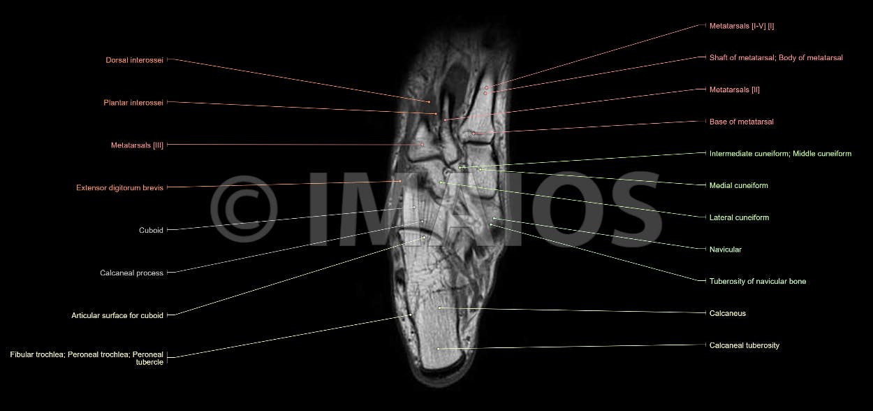
Anatomy of the foot and ankle - MRI from www.imaios.com It arises from the base of the fifth. The paraspinal muscles, which are innervated by the spinal nerve dorsal ramus, are also frequently tested. The aim of this review is to provide the reader with a comprehensive overview of the magnetic resonance imaging (mri) characteristics of the most common benign and malignant soft tissue neoplasms which occur around the foot and ankle. The muscles acting on the foot can be divided into two distinct groups; • muscle edema is seen secondary to multiple etiologies including trauma, infectious and inflammatory processes, autoimmune disorders, neoplasms, and denervation injuries • on mri muscle edema is characterized by increase in free water within the muscle • muscle edema is seen on mri as increased signal on fluid sensitive sequences t2 fs Mri of the ankle and feet 12 photos of the foot muscle anatomy mri.magnetic resonance imaging (mri) is the modality of choice in diagnosing accessory muscles, delineating their relationship to adjacent structures, and differentiating them from soft tissue tumors. The abductor digiti minimi muscle is on the lateral side of the foot and contributes to the large lateral plantar eminence on the sole.
Hip pelvis thigh knee lower extremity/shin ankle foot.
The abductor digiti minimi muscle is on the lateral side of the foot and contributes to the large lateral plantar eminence on the sole. Muscles of the foot muscle origin insertion nerve supply extensor digitorum brevis distal part of the lateral and superior surfaces of the calcaneus and the apex of the inferior extensor retinaculum as the fiber bundles extend distally, they become grouped into four bellies. They are mainly responsible for assisting some of the extrinsic muscles in their actions. Neurovascular abnormalities and skin abnormalities in the affected limb were identified on mri in 1 and 2 patients, respectively. This small, thin muscle is absent in about. Coronal images are perpendicular to the long axis of the metatarsals. Routine ankle magnetic resonance imaging (mri) tests involve taking images of the foot and ankle in the axial, coronal, and sagittal planes parallel to the tabletop(2). The muscles acting on the foot can be divided into two distinct groups; The muscle that removes the big toe (m.abductor hallucis) lies superficially along the medial edge of the foot. Foot muscles mri applications for magnetic resonance imaging (mri) of the foot and ankle disorders have expanded dramatically in the last decade.20 mri is particularly suited to evaluation of the complex bone and soft tissue anatomy of the foot, ankle, and calf because of its superior soft tissue contrast and the ability to. Mri of the ankle and feet Foot ulceration can subsequently lead to infections, such as cellulitis and osteomyelitis, and this may eventually the mri examination includes special attention for positioning of the foot. Specific force for toe flexor muscles in this study was somewhat smaller than these values.


0 Comments